How to stretch your pectoral muscles. Expansion of the chest. Stretch against the wall
We are always frightened by pain in the heart area, since their occurrence is often associated with myocardial infarction, the mortality rate from which is very high...
Problem: pain in the heart area
We are always frightened by pain in the heart area, since their occurrence is often associated with myocardial infarction, the mortality rate from which is very high.
However, in some cases these fears are unfounded.
Pain can be caused by other reasons, but only a doctor can find out.
One of the most common causes of such pain is a painful spasm of the pectoralis major and minor muscles.
Rice. 53. Pain in the heart area
When they are affected on the right side chest Pains similar to pain in the heart may appear.
Many patients describe the pain as dull, aching, or boring.
They occur during physical stress:push-ups, pull-ups, lifting and carrying loads, sharply moving the arm back when swinging while playing volleyball, tennis, badminton.
Due to increased pain, the movement of the arm back is usually limited.
In addition, pain can appear at rest, and sometimes at night, disturbing sleep.
In addition, pain appears in the subclavian region, the anterior surface of the shoulder and spreads to the arm, accompanied by numbness and weakness of the arm muscles.
This is explained by compression of the neurovascular bundle under the tense pectoralis minor muscle.
Few people know that when the pectoral muscles are damaged, periodic rhythm disturbances may occur without other signs of heart damage.
Main functions of the pectoral muscles:
Adduct and internally rotate the shoulder;
Raise the ribs with a fixed shoulder blade or arm during take a deep breath, taking an auxiliary part in breathing;
Pull the shoulder blade forward and downward.
Causes of spasm:
Osteocondritis of the spine;
Lifting and holding heavy objects in front of you without warming up;
Abrupt withdrawal of the arm when throwing a ball, serving in tennis, etc.;
Being in a draft and overcooling tired muscles;
Quick push-ups from the floor, pull-ups on the horizontal bar, pushing a barbell without prior warm-up;
Overstrain of the adductor muscles of the arms during prolonged use with a saw, wire cutters, etc.;
Persistent cough;
Prolonged compression of muscles by the strap of a heavy backpack;
Injury to the pectoral muscles;
Muscle strain when sitting for long periods of time with drooping shoulders.
How to find the affected muscle
Palpation of the pectoral muscles is carried out with the opposite hand. Typically, the site of maximum pain corresponds to the site of maximum spasm.
If you have difficulty, then moving your arm back slightly, which causes tension in the pectoral muscles, can make the search easier.
In this position, the spasmodic area becomes more sensitive to pressure and is clearly felt in the form of a compaction or cord.
As a rule, finding painful areas does not cause difficulties.
Stretching is carried out in 2 positions.
Position No. 1 (Fig. 54)
Rice. 54. Stretching the pectoralis major and minor muscles (position No. 1): a - clavicular portion; b - sternal portion; c - costal and abdominal portions
Sitting on a chair near the doorway, move the arm on the affected side to the side, bend it at the elbow, the hand and forearm should be adjacent to the doorway from behind.
Shifting the torso forward and turning it to the healthy side increases the amplitude of the stretch.
Stretching:
a) the upper part (clavicular portion of the pectoral muscles) facilitates the lowering of the shoulder below the horizontal level;
b) middle part (thoracic portion) - position of the shoulder at a horizontal level;
c) lower part (costal and abdominal portions) - raising the shoulder above the horizontal level.
Duration of stretching is 5-10 minutes.
Perform it daily until the pain on palpation completely disappears.
Position No. 2 (Fig. 55)
Lie on your back with your head on a pillow, move your arm to the side, bend it at an angle of 90° at the elbow joint, and cover it with a pillow on top.
The amplitude of the stretch is regulated by turning the body in the healthy direction.
Rice. 55. Stretching the pectoralis major and minor muscles (position No. 2)
Stretching the upper, middle and lower portions of the pectoral muscles is facilitated by abducting the shoulder towards the legs or head, similar to the stretch in position No. 1.
Prevention measures:
Perform comprehensive treatment of osteochondrosis in a timely manner;
Be sure to warm up before upcoming work involving heavy lifting and sports;
Avoid hypothermia and drafts;
When wearing a backpack, move its strap to the edge of the shoulder, and so that it does not put too much pressure, place a wide pad under it;
When sitting for a long time, create support for your elbows or use a chair with comfortable armrests;
Take timely breaks from work, rationalize your activities so that there are no conditions for overworking the adductor muscles of the arms. published .
Leontyev A.V.
If you have any questions, please ask
P.S. And remember, just by changing your consciousness, we are changing the world together! © econet
A pectoral muscle strain is a type of injury that occurs due to excessive stress on it. It can happen on both the right and left side of the chest as a result of performing physical exercise or even while coughing.
Symptoms
Symptoms of a muscle strain can vary depending on the underlying cause. Some may be so severe that you will be unable to move or perform daily activities. The most common symptoms of a pectoral muscle strain are:
- Stiffness and pain, especially if the sprain was caused by severe coughing
- Sharp pain at the site of muscle strain when coughing or sneezing
- Difficulty moving
- Pain when trying to bend or turn if the muscles of the back and abdomen are also affected
- Labored breathing
- In some cases, pain and tenderness when touching the affected area
Often, a person is most frightened by chest pain, because pain can be a sign of life-threatening diseases, such as a heart attack, pulmonary hypertension, or pulmonary embolism. But in fact, more often these sensations are associated with non-dangerous conditions, including muscle strains, the effects of coughing, heartburn, or simply minor injuries. Although respiratory and cardiac diseases cannot be ruled out, a visit to the doctor may be necessary.
How does stretching occur?
The human chest consists mainly of two muscles: the pectoralis major and the pectoralis minor. The first is the largest muscle in this area, while the second is small and located below it. These muscles are connected by the breastbone, also known as the sternum. Their functions are to control the movements of the arms and shoulders. During these actions, any of the muscles can be injured.
A strained chest muscle can occur as a result of simple activities such as heavy lifting, improper exercise, or severe coughing.
This can lead to stiffness and pain, which often varies depending on the extent of the muscle damage. With a mild injury, the pain is usually not severe and goes away within a couple of days. However, in a more severe case, the muscle tear may be so severe that it limits your movement and breathing.
Both men and women are at risk for pectoral muscle strains. Most injuries occur from sudden or severe strain. Anatomy female breast different from men's. In women it has more high level contains fatty tissues that help protect the area. They help protect women's mammary glands and ducts. This does not mean that a woman cannot stretch her chest muscles, just like men, they can also get the same injuries.
How not to confuse it with a heart attack?
Sometimes it can be difficult to differentiate between a chest muscle strain and a heart attack. According to experts, an urgent and proper medical examination is required, which can last more than a day. Symptoms of a heart attack include:
- Severe pain radiating to the left arm
- Shortness of breath accompanied by chest discomfort
- Dizziness
- Nausea and cold sweat
Not all people who have a heart attack will experience the above symptoms. For some, they may appear suddenly or appear within a few days. If you experience any unexplained pain in the chest area, you should consult a doctor as soon as possible.
How long does it take to treat?
Healing time may vary depending on the severity of the injury.
The recovery period for the chest muscles may be as follows:
- 7 – 10 days for class I muscle injuries
- 2 – 5 weeks for grade II muscle injuries
- 10 - 12 weeks for grade III (most severe)
Strained pectoral muscle from coughing
Coughing is a common cause of chest muscle strain. It is a periodic protective reflex that helps clear the airways of liquids, irritants, and other foreign particles.
A cough consists of three phases: inhalation, forced exhalation with a closed glottis, and a sharp release of air from the lungs. To perform the second and third of these processes, the muscles become very tense.
A severe or prolonged cough can cause straining and straining. various muscles chest wall, cause muscle fibers to tear, causing pain, stiffness and tenderness in the chest. Although it can be very painful, this type of pectoral muscle strain is not considered serious. For most people, overall improvement occurs within a day or two.
Stretching the pectoral and back muscles
The pectoral and upper back muscles cover the upper torso area. They start from the bottom of the neck and end at the beginning of the abdominal cavity, and also include the muscles of the shoulders.
These muscles are responsible for controlling movement, which includes moving the arms and head. They also control other vital activities such as breathing.
Most of the functions of the chest are controlled by the pectoralis major muscles. These are fan-shaped muscles that make up the bulk of the chest muscles.
Often, several different muscles of the back and chest can be affected by a sprain at the same time, or so it may seem due to the spread of pain. Causes of damage:
- A herniated disc in the thoracic spine can cause spasms, numbness, and weakness in the back muscles. This is a common cause of pain between the shoulder blades on one or both sides, especially after exercise.
- A spinal sprain is a tear in the muscle tissue around the facet joints caused by overuse.
- A costovertebral joint sprain caused by damage to the connective tissue of one or more joints located near the spine where the ribs attach to the vertebrae. This can be caused by prolonged lying, flexion or extension.
Pain and difficulty breathing when the pectoral muscles are stretched
Injuries or injuries sometimes cause muscles in the chest and costovertebral joint to spasm, which can interfere with breathing, coughing, or sneezing.
Sprains of the costovertebral joint usually occur after a sudden movement involving rotation, bending or extension of the spine. This joint consists of the end of the rib and the flat section of the thoracic vertebra. Straining the area when moving or doing certain exercises can damage the ligaments or cartilage, causing pain when breathing.
Left side stretch
A muscle strain in the chest area can occur on both sides at the same time or on just one side. A sprain on the left side can be caused by injury, dislocation of the facet joints, or damage to the ligaments of the costovertebral joints on the left side of the chest.
Regardless of the cause, pain on this side requires urgent medical diagnosis to rule out the possibility of serious, life-threatening diseases such as heart attack and others.
Treating the underlying cause of a muscle strain is the best way to relieve and prevent future complications.
Right side stretch
Just like on the left side of the chest, a muscle strain on the right can happen as a result of a cough, injury, bruise or sprain in this area.
In mild cases of muscle strain, simple remedies such as RICE therapy (rest, cold compress, compression and elevation of the injured area) can help relieve pain, stiffness and chest discomfort caused by muscle damage. However, urgent medical examination is recommended. Especially when the symptoms do not go away within one day.
Most people associate chest pain with serious medical conditions such as a heart attack, pulmonary hypertension, or thromboembolism. This is not always true, but this option cannot be ruled out; a correct medical diagnosis is required.
Treatment
Treatment for a pectoral muscle strain often depends on the underlying cause. If it is due to a strong cough, which is the main cause, then the problem is not serious. However, it can be unpleasant and very painful.
If cough is the root cause, then its treatment is The best way to relieve pain. When the severe cough stops, the chest muscles will have time to rest and heal. Microscopic tears in the chest muscles will go away on their own if they are not stressed.
For a mild cough, simple home remedies can help, for example, take a tablespoon of basil juice or aloe tincture with honey 3 times a day. In severe cases, your doctor may prescribe some cough suppressants or expectorants to make coughing easier.
Other treatment options for a muscle strain include:
- Anti-inflammatory and pain relievers to relieve pain and stiffness caused by muscle tension. These are over-the-counter ointments and tablets that a pharmacist will offer at any pharmacy.
- Cold and warm compress. The first is to relieve inflammation and the second is to speed up healing
- Ultrasound therapy can also be used to provide effective treatment. It promotes blood supply to damaged chest muscles
- Severe cases of chest muscle strain may require surgery.
Small pectoral muscle- “smaller sister” of the more massive and well-known pectoralis major muscle. However, even a muscle this small can cause serious problems.
It is located under the pectoralis major muscle, originates from the 3rd, 4th and 5th ribs, passes laterally upward and is attached to the coracoid process of the scapula. Functions of the pectoralis minor muscle: stabilization of the scapula, as well as its lowering, retraction and downward rotation.
When the scapula is in a fixed position, this muscle also takes part in lifting the chest. Having explained the functions of the pectoralis minor, estet-portal.com will tell you what problems may be associated with it.
Condition of the pectoralis minor muscle and postural disorders
The pectoralis minor muscle is a fairly significant factor in the formation of correct (or incorrect) posture. One of the functions of the pectoralis minor is to retract the scapula—moving it forward, away from the spine, and around the rib cage, resulting in a hunched position in the shoulders.
Unfortunately, the lives of most of us are dominated by a sedentary lifestyle - we spend the lion's share of time at computers, driving, leaning over the desk in unnatural positions, when our shoulders are rounded and tilted forward, our backs are hunched. Over time, this position inevitably leads to overstrain of the pectoralis minor muscle (the appearance of muscle knots, or clamps).
Poor posture, although a big one, is not the only problem caused by overstrain of the pectoralis minor muscle. Therefore, below estet-portal.com will consider:
- what symptoms and problems appear with overexertion of the pectoralis minor;
- what is pectoralis minor syndrome?
- how to get rid of pain caused by overexertion of the pectoralis minor muscle.
Overstrain of the pectoralis minor muscle: the starting point of serious disorders
An overused pectoralis minor muscle significantly affects the function of the shoulder joint and increases the risk of injury.
Perhaps the most common problem associated with the pectoralis minor is subacromial impingement syndrome.
In this case, the lack of space in the area between the humerus and the acromion leads to pinching of the tendons of the supraspinatus muscle and subacromial bursa, especially when raising the arm above the shoulder, as well as during rotational movements.
Attention!
This lack of space may be directly related to a tight pectoralis minor muscle due to anterior tilt of the scapula and decreased subacromial arch.
In this case, patients complain of:
- dull pain in the shoulder;
- increased pain when raising the arm up;
- problems sleeping as a result of pain, especially when lying on the affected side;
- a characteristic crunching or clicking sound when lowering your hand;
- limitation of joint mobility;
- weakness in the hand.
In addition to increasing the risk of entrapment, an overtightened pectoralis minor muscle, together with the serratus anterior muscle, moves the glenoid fossa of the scapula into a more vertical position, which leads to increased abduction, rotation and winging of the scapula.
Due to changes in the position of the scapula, the levator scapulae muscle and superior fibers trapezius muscle begin to strain intensely in an attempt to stabilize her position. Ultimately, this leads to overstrain of the neck and shoulder muscles and the appearance of many trigger points in this area, which is typical for many who work at a desk (or on the couch, with a laptop on their lap).
Pectoralis minor syndrome - when nerves and blood vessels are affected
In 50% of cases, it is the pectoralis minor muscle that is the root cause of thoracic outlet compression syndrome (or simply pectoralis minor syndrome). What it is?
Pectoralis minor syndrome is manifested by compression of the nerves and blood vessels located in the anterior part of the shoulder and chest - the brachial plexus, subclavian artery and vein. Pinched nerves and blood vessels necessarily make themselves felt, causing significant physical discomfort.
Symptoms of this syndrome vary depending on the location and degree of compression of the neurovascular bundle and can manifest as:
- pain throughout the entire arm - from the shoulder to the little finger;
- numbness, decreased sensitivity, tingling in the limbs;
- weakness in the hand;
- pallor of the upper limb;
- decreased hand temperature;
- increased pain during movement.
As you can see, the pectoralis minor muscle, or more precisely, its overtension, can cause problems affecting the area of the shoulder joint, neck and arm. But this is only part of the whole picture: as a result of muscular compensation, which passes through the kinetic chain, the negative impact of overexertion of the pectoralis minor muscle can affect even bottom part back and pelvic area.
How to relax the pectoralis minor muscle: stretching and trigger point massage
The figures below show the location of the trigger points of the pectoralis minor muscle and a diagram of the reflected pain when pressing these points:
Fortunately, the pectoralis minor responds well to therapy such as massage and/or stretching. However, it is worth noting that it is not so easy to cope with an overstrained pectoralis minor muscle on your own, so estet-portal.com recommends either contacting a specialist (which, of course, is better) or asking a friend/spouse/relative for help.
Stretches to eliminate pectoralis minor trigger points:
- Lie down on a cylindrical object (a special cushion or a tightly rolled blanket or towel) so that it is in line with the spine. In this position, lower your shoulders down towards the floor, relax, feel the relaxation in the front of your shoulder and upper chest. In this case, your arms can first be placed along the body, and then spread at an angle of 80, 120 and 160 degrees.
- Sit on the floor and ask an assistant to stand behind you. Bring both arms back and ask the assistant to gently, smoothly and with little force move the arms further.
Massage of trigger points of the pectoralis minor muscle:
To massage the pectoralis minor muscle, it is better to contact a specialist who can correctly determine the location of trigger points and effectively influence them. At home, massage can be performed using a tennis ball in a lying position (placing the ball on the floor) or standing (clutching the ball between the wall and the pectoralis minor muscle):
In addition to stretching and massage, try doing exercises aimed at strengthening the antagonist muscles of the pectoralis minor muscle. Focus on the middle and lower trapezius, rhomboids, serratus anterior, infraspinatus, and dorsal deltoids.
Source: https://estet-portal.com/statyi/malaya-grudnaya-myshtsa
Pectoral muscles
| Deep push-ups Description Deep push-ups. Initial position. When using stops, the exercise becomes more complicated due to... | Bench pressDescription Bench press. Initial position. Lying in the exercise machine, feet resting on the floor. The head lies on... | Inward chest press on a block Description Inward chest press on a block. Initial position. Standing in the center of the blocks, one leg bent at the knees... |
| Dumbbell bench press Description Dumbbell bench press. Initial position. Lying down horizontal bench, feet resting on the floor. Head… | Dumbbell Bench Press (Negative Incline Bench)Description Dumbbell Bench Press (Negative Incline Bench) | Dumbbell bench press (bench with positive incline)Description Dumbbell bench press (bench with positive incline). Initial position. Lying on incline bench, legs… |
| Dumbbell bench press with palms facing inward Description Dumbbell bench press with palms facing inward. Initial position. Lying on a bench, feet flat on the floor. Hands with... | Dumbbell bench press with palms inward (negative incline bench) Description Dumbbell bench press with palms inward (negative incline bench). Initial position. Lying on... | Dumbbell bench press with palms inward (bench with positive incline) Description Dumbbell press with palms inward (bench with positive incline). Initial position. Lying on a bench with... |
| Dumbbell bench press on the floor Description Dumbbell bench press on the floor. Initial position. Lying on the floor, your legs can be bent at the knees to... | Dumbbell bench press on a fitballDescription Dumbbell bench press on a fitball | One-arm dumbbell bench press Description One-arm dumbbell bench press. Initial position. Lying on a horizontal bench, feet resting on the floor... |
| Smith Machine Bench PressDescription Smith Machine Bench Press. Initial position. Lying on a horizontal bench in Smith's car, legs... | Smith Machine Bench Press (Negative Incline Bench)Description Smith Machine Bench Press (Negative Incline Bench). Initial position. Lying down in Smith... | Smith Machine Bench Press (Positive Incline Bench)Description Smith Machine Bench Press (Positive Incline Bench). Initial position. Lying on a bench in... |
| Wide Grip Smith Machine Bench Press Description Wide Grip Smith Machine Bench Press. Initial position. Lying on a horizontal bench, legs... | Bench press reverse grip Bench press with reverse grip Description Bench press with reverse grip. Initial position. Lying on a horizontal... | Reverse Grip Bench Press (Positive Incline Bench) Description Reverse Grip Bench Press (Positive Incline Bench). Initial position. Lying on a bench with... |
| Bench press with a wide reverse grip Description Bench press with a wide reverse grip. Initial position. Lying on a horizontal bench, feet resting on... | Wide grip bench press Description Wide grip bench press. Initial position. Lying on a horizontal bench, feet resting on the floor.... | Chest press on a block Description Chest press on a block. Initial position. Standing in the center of the block frame. We bend one leg slightly in... |
| Bench press This is a basic exercise for the pectoral muscles. Starting position Lying on a horizontal bench. The most stable stop... | Bench press (negative incline bench)Description Bench press (negative incline bench). Initial position. Lying on a bench with your head down... | Bench press (bench with positive incline)Description Bench press (bench with positive incline). Initial position. Lying on an inclined bench, emphasis... |
| Bench press lying on the floor Description Bench press lying on the floor. Initial position. Lying on the floor, for greater stability, you can... | Bench press on the neck Description Bench press on the neck | Reverse Dumbbell Bench Press Description Reverse Dumbbell Bench Press. Initial position. Lying on a horizontal bench, hands with dumbbells... |
| Push-ups Description Push-ups. Initial position. Standing in a plank position, with your toes pointing to the floor. The arms are straight, positioned... | Dips (chest) Description Dips (chest) | Dips with weights Description Dips with weights. Initial position. On uneven bars with straight arms. The body is tilted... |
Source: http://bodybuilder.ucoz.ru/index/uprazhnenija_dlja_razvitija_grudnykh_myshc/0-43
Pectoral muscles: location and functions:
The muscles located in the thoracic region of the human torso perform a lot of voluntary and involuntary functions. Let's consider their diversity, location and main tasks.
Pectoral muscles
IN human body four types of muscles of the pectoral region:
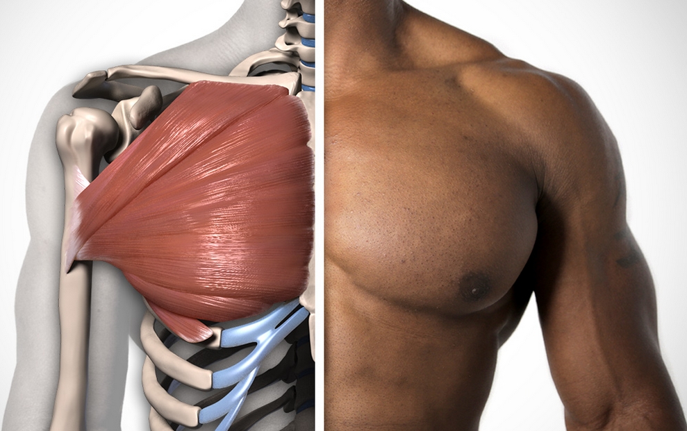
- The pectoralis major muscle is the most visible. This is the muscle mass of the chest that shapes its appearance. It helps with breathing, brings the arm to the body, and is responsible for turning it inward.
- The pectoralis minor muscle is not visually visible, because it is located behind the major muscle. It is involved in the breathing process, and it is also what pulls the shoulder blades forward and down.
- The subclavius muscle, located between the uppermost rib and the collarbone, moves the collarbone inward and downward, strengthens the sternoclavicular joint, and elevates the first pair of ribs.
- Front serratus muscle located on the side of the chest. In terms of its location, it is the lowest pectoral muscle. Together with the rhomboid, it forms a fairly powerful muscle mass that covers the entire human torso and presses the scapula to it.
Proprietary muscles of the sternum
Don’t forget about your own chest muscles:
- Aperture - main muscle during the breathing process. This is the muscle-tendon septum between the thoracic and abdominal regions. It helps to increase intra-abdominal pressure, contracting together with the abdominal muscles.
- Subcostal muscles involved in the act of inhalation. Located on the dorsal surfaces of the lower pairs of ribs. Their bundles seem to be thrown over one rib.
- The external and internal intercostal muscles are participants in the “inhalation-exhalation” process. Their beginning is different costal edges.
Pectoralis major muscle: location
The paired wide Pectoralis major muscle is located in the anterosuperior region of the chest. Involves the shoulder joint. Its insertion point is the flat tendon of the intertubercular groove of the humerus. Muscle location:
- The upper part (another name is the clavicular) - from the medial line to the anterior clavicular surface. Here it forms the clavicular fossa and the deltoid-pectoral groove.
- Next, the middle part of the muscle is observed - the sternocostal muscle. It begins at the anterior surface of the sternum and ends in the area of the 2-7th pair of ribs.
- The lowest and most weakly expressed is the abdominal one. It starts from the anterior vaginal wall of the rectus abdominis muscles.
Functions of the pectoralis major muscle
This pectoral muscle performs the following tasks:
- adduction and internal rotation of the shoulder;
- bringing the arm raised horizontally into a sagittal position (left or right);
- pronation - inward rotation of the limb;
- arm flexion;
- helping to expand the chest during the breathing process.
Location of the sternum minor muscle
A flat, triangular-shaped pair of Pectoralis minor muscle is located directly under the major pectoral muscles. It is attached to the coracoid process of the scapula by a short tendon. The pectoralis minor muscle controls the joints from the shoulder blade to the ribs. The location of this muscle component in the human body:
- The beginning is individual teeth in the area of the 2-5th pair of ribs, in close proximity to the junction of their bone and cartilage tissue.
- Next, the muscle stretches upward in the lateral direction. At the same time, its bundles converge.
Functions of the pectoralis minor muscle
Tasks performed by the sternum minor muscle:
- movement of the scapula up and down;
- abduction (reduction) of the shoulder blades;
- auxiliary function during breathing - with a stably strengthened scapula, the muscle raises the ribs in this process.
Location of the serratus anterior muscle
The wide flat paired Serratus anterior muscle is located in the anterolateral region of the sternum. Her top part, like the pectoralis minor muscle, is hidden under the sternum major muscle. The lower one is superficial, located under the pectoral fascia.
Together, the serratus anterior muscles protect the outer costal surface and then extend under the scapula. Here it is attached to the lower corner of the scapular triangle along its medial edge.
Here, it should be noted, the most developed bundles of this muscle are located. Location - 2 starts:
- The outer surface of the 8-9th pair of ribs (8-9 muscle teeth).
- From the tendon arch passing between the 1st-11th pairs of ribs.
Functions of the serratus anterior muscle
This pectoral muscle performs the following set of functions:
- pulling the shoulder blades away from the spinal column;
- lateral (side) displacement of the lower angle of the scapula;
- rotation of the scapula along the sagittal axis;
- fixing the position of the scapula, connecting it to the chest (in tandem with the rhomboid muscle);
- auxiliary respiratory function (during inhalation) - with the condition that the belt of the upper limbs remains motionless.
Location and functions of the subclavius muscle
The small oblong paired Subclavius muscle is almost parallel to the collarbone, located slightly below it. Its surface is hidden by the extensive large muscle of the sternum. In the lateral and vertical direction, this muscle is attached to the inferior acromial clavicular region. The subclavius muscle performs the following functions and tasks in the body:
- movement of the clavicle down and towards the medial line;
- raising the upper pair of ribs;
- holding the collarbone in the sternoclavicular joint;
- auxiliary function during the respiratory process;
Thus, all four types of paired pectoral muscles (major, minor, serratus anterior and subclavian) perform a large range of functions - various movements of the limbs, shoulder blades, collarbone, participation in the respiratory process. The quality of performance of arbitrary tasks, appearance (relates to the pectoralis major muscle) depend on the sports behavior of a particular person.
Good afternoon, honest people, take the faces out of your beards, tea, we are not having a memorial service, but quite the opposite! This Friday, oddly enough, we will pay attention to the male audience of the project, because our ladies are already all beautiful, and we will pull our brother up :). The topic of the note is how to expand the chest. And this is exactly what you will learn from it, or rather, we will look at the anatomy respiratory system, let's find out how to breathe correctly during strength exercises and, of course, we will analyze a specific training program aimed at voluminous breast enlargement.
So, guys, send everyone to the nearest 10 minutes, because there is a serious men's conversation ahead.
How to expand the chest? FAQ questions and answers
If you follow the news of the project and the release of fresh articles, then you are aware that we have just completed a women’s series of narrow notes aimed at correcting the figure. It may seem to the male part of the audience that we are somehow depriving the stronger sex of attention, this may be true, but it’s all due to lack of initiative on your part. The ladies simply bombarded their mail and comments with questions that worried them and got what they wanted - a series of materials with specific training programs for correcting problem areas.
With regard to our brother, one gets the impression that everything is top-notch with them (us), and if there’s something that worries us globally, it’s either gaining muscle mass or the “animal issue,” i.e. how to remove it. Well, if that’s the case, then we don’t really bother ourselves with articles on men’s topics. If things continue like this, then we will completely become a feminine project with a name like the ABC of Fitness or Fitolyazhka :). Well, let’s not talk about sad things, but let’s touch on what I’m sure is relevant for many novice athletes (especially ectomorphs) topic called how to expand the chest?
Do you know that not only the butt is flat, but also the chest? And if the “meat” on the buttocks can be increased, then with the chest the situation is somewhat more complicated. And the most important thing when converting a plane into volume is time, in the sense of starting on time. Often this time is missed and a young man, having crossed a certain young age and entered adulthood, remains with an undeveloped and sunken sternum. All subsequent attempts to “inflate” the chest bring extremely insignificant results. Therefore, if you are a beginner (recently arrived at Gym) age up to 25 (better 20 ) years and at the moment your priority is to have sculpted abs or big biceps, then stop toiling around with bullshit :) and start developing the volume/depth of the chest. Cubic abs and other desires can easily wait, but breasts cannot.
Well, have you already quit? Then we move on to the theoretical and practical side of the issue. Go!
Note:
For better assimilation of the material, all further narration will be divided into subchapters.
Human respiratory system: questions of anatomy
The respiratory system provides the body's cells with oxygen, removing carbon dioxide and waste products that accumulate in the human body over time. DS helps the body in the exchange of gases between air and blood, between blood and billions of cells. In addition to air distribution and gas exchange, the breathing filter system warms and humidifies the air we breathe.
It is customary to highlight 3 main parts/components of the DS:
- Airways. Includes the nose, mouth, pharynx, larynx, trachea, bronchi and bronchioles. They carry air between the lungs and to the external environment (outside the body);
- Lungs. Act as functional units of the respiratory system, by passing oxygen into the body and removing carbon dioxide from the body;
- Breathing muscles (including diaphragm and intercostal muscles)- work together to act as a pump, pushing air in and out of the lungs during breathing.
In the note key, we are most interested in the respiratory muscles, so we will pay closer attention to them.
The main breathing muscle in the human body is the diaphragm - a thin layer skeletal muscles, which forms the “floor” in the chest. When the diaphragm contracts/compresses, it moves (several centimeters) down into the abdominal cavity, expanding the space inside the chest cavity and pumping/blowing air into the lungs. During the process of relaxation of the diaphragm (exhalation), the volume of the chest decreases, the lungs compress, and the pressure in them increases (becomes above atmospheric), and the air rushes out through the airways.
Visually it all looks like this.

Between the ribs there are many small intercostal muscles that help the diaphragm with the expansion and contraction of the lungs. These muscles are divided into 2 groups:
- internal intercostal muscles (exhalatory muscles) - a deep set of muscles that are involved in the act of forced exhalation;
- External intercostal muscles (inspiratory muscles) are deep chest muscles that raise the ribs and expand the chest.
What is the process of breathing?
When you take a deep breath, the diaphragm contracts and moves downward. This action opens up more space in the chest cavity, allowing the lungs to expand. The air travels down the trachea and into the bronchi, ending up in air sacs called alveoli.
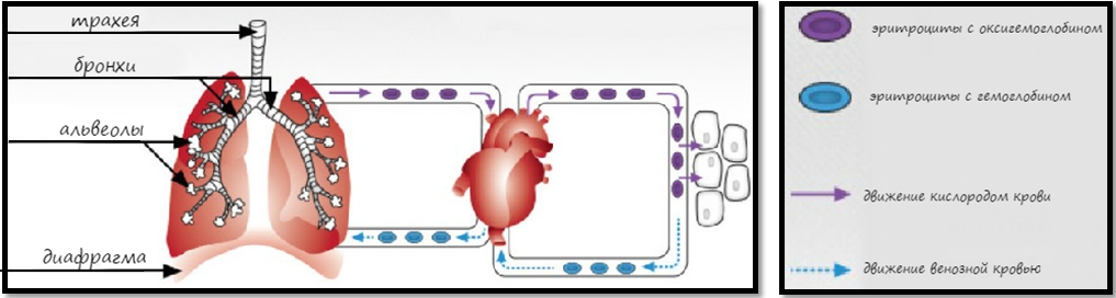
Oxygen then passes through nearby capillaries into the alveoli, where hemoglobin, a red blood cell protein, moves it into the blood. Oxygenated blood is carried through the pulmonary vein on the left side of the heart and then pumped to the rest of the body's tissues. Once this happens, carbon dioxide begins to move through the capillaries into the alveoli, traveling to the right side of the heart through the pulmonary artery.
As for exhalation, during the process the diaphragm relaxes and moves up into the chest cavity. The intercostal muscles inside the ribs relax, which also leads to “shrinkage” of the chest cavity. During exercises (for example,), the abdominal muscles contract more often, pushing the diaphragm against the lungs more often. When this happens, carbon dioxide is expelled faster, increasing the frequency of your breaths.
How an athlete can breathe during strength exercises
Technique No. 1.
Correct/timely delivery of oxygen to the “starving” muscles from the Volga region during heavy basic exercises allows you to increase the athlete’s performance and increase the duration of the workout.
When moving to any serious weights (for example, in deadlift) Often the athlete loses his breath, and because of this he finishes the exercise earlier than possible. The result of this situation is a reduction in the training volume of the load and underloading of the target muscle groups.
Therefore, it is advisable, as you move towards heavier weights, to use the Valsalva breathing technique (Valsalva maneuver, VM). This is the process of maximal inhalation while maintaining pressure. The most significant example of such a “maneuver” is the process of sitting in the restroom and helping (by inhaling, tensing and pushing) to your intestines in removing the products of your own vital activity.
This breathing technique adds stability to the spine and increases the “drive” from the legs to the arms and then to the bar.
As a result of deep inhalation and air retention, intra-abdominal pressure increases, which ultimately creates a more “stable frame” and avoids injury. Another benefit of the Valsalva breathing technique is that it increases blood pressure, which could theoretically improve the delivery of nutrients to cells.
Note:
If the athlete has problems with blood pressure (for example, hypertensive), then it is better for him not to use the technique of maximum inhalation while holding pressure. Also, such an athlete should not perform more than 1-2 heavy basic exercises from the “golden three”. People who do not suffer from pressure surges can use the VM technique and the number of exercises from 2 to 3 per workout (but not more 1 once a week).
There is one subtlety in the Valsalva breathing technique, or rather, by-effect- holding your breath for a long time during the exercise can lead to loss of consciousness. Therefore, the rule when working with VM is to hold your breath only until you pass the dead point of the deadlift/press/squat, then exhale.
Technique No. 2.
The above breathing technique can be used by an athlete of average fitness level, but for a beginner, the optimal exhalation pattern code-named “teapot” :). It consists in the fact that in the process of performing an exercise (for example,), the athlete takes a breath of medium depth and then slowly and through pursed lips (lips with a tube) exhales at the heaviest phase (force/projectile ejection). This exhalation is similar to steam coming out of a heated kettle, and it allows you to use some of the stabilizing properties of the inhalation without creating a large surge in pressure.
Proper breathing as one of the “tools” for expanding the chest and gaining muscle mass
Above we looked at 2 different techniques breathing. Which one is better to use for gaining muscle mass, and can breathing really affect muscle volume?
Scientists and sports medicine specialists have found (data from MUSCLE & FITNESS No. 7-8 2005 ) , What correct breathing during strength exercises helps to increase mass. Rhythmic breathing is one of the main conditions for anabolism. Its blockage leads to a weakening of blood flow in the working muscle. Moreover, when breathing is blocked (blocking inhalation) the athlete's body is deprived of oxygen - an important agent muscle growth. The more oxygen in the blood, the better the conditions for muscle growth.
Therefore, breathing strategies during training may include the following:
- rhythmic (classical-bodybuilding)- when working with basic exercises (conditionally basic, for example, military chest press, dumbbell flyes/presses);
- breathing using the Valsalva technique – when working with heavy basic exercises (bench press, deadlift, squats).
According to sports medicine specialists, the correct rhythmic breathing pattern, using the example of the military chest press, consists of: 6 stages and looks like this:
- remove the bar from the racks;
- holding it on straight arms, inhale deeper than usual;
- hold your breath and lower the barbell to your chest (until light touch);
- press the barbell upward, still holding your breath;
- When you pass the hardest point of the climb, exhale.
- when the bar is in the IP, inhale again.
Use different breathing techniques depending on the intended workout and the exercises involved.
Actually, we have laid down some construction and “breathing” theories; now we will deal with the issues of transferring the chest from plane to space.
How to expand the chest: the practical side of the issue
If you take a quick look at your gym, you will draw the following conclusions about the goals of the trainees, namely: young ladies want to lose weight and become smaller, guys need to enlarge or lengthen everything :). And because The pectoral muscles are one of the largest muscle groups, so increasing their volume will make a significant contribution to improving a person’s composition.
Under the expression “expand the chest” (in the context of this note) we will understand the following:
- increase in chest width (horizontal linear dimension);
- adding “depth” (increase vertical size);
- adding volume (increase in girth size);
- increase in muscle mass.
All this is achieved through a special training program aimed at “pulling out” the breasts by influencing anatomical features(including) pectoral. What they are, we will find out further in the text.
“Tools” for chest expansion
First of all, take a crowbar and an expander and…
To expand the chest, the athlete must exert intense pressure on 3 component:
- bones;
- muscles;
- connective tissue.
Let's go through each “tool” separately and start with...
I. Expansion of the skeleton
Did you know that bone completely remodels itself every three months? This suggests that if time is not lost (the athlete is young, up to 25 years old, and his growth zones are still open), then you can “grow” your bones, i.e. conditionally from 15 see wrist do 17,5-18 . Bones react to weight, i.e. their density increases as the weight used by the athlete increases. Thus, the training strategy for a beginner who has decided to “bone” increase should be a gradual transition to basic movements, strengthening ligaments/tendons and increasing the weight of the weight, rather than swinging the press on a bench and doing dumbbell bicep curls.
To stimulate your skeletal system to grow, use the following “tricks”/exercises:
No. 1. Holding your breath
Follow next exercise for some long time ( 3 month) every morning:
- clasp your hands in front of your waist and take as deep a breath as possible;
- hold your breath while raising your clasped hands up to a position in front of you;
- while holding your breath, try to move your arms to the sides, overcoming the resistance of your clasped hands;
- stay in this “extended” position for as long as you can;
- repeat 15 once.
No. 2. Breathing squats
The exercise is as follows: classic squats are performed with a barbell on the shoulders according to the following scheme:
- done before squats begin 3 large inhalations;
- after the 3rd inhalation, the breath is held and a squat with the barbell is performed to parallel with a quick “rise to the top”;
- after straightening, exhale strongly;
- the pattern is repeated;
- Total 3 approach 15-20 repetitions.
II. Working with connective tissue
Here pullovers with a dumbbell/barbell lying across the bench will help us/you. This great exercise to strengthen and stretch the pectoral connective tissue.

III. Work on all 3 sections of the pectoral muscles
The total development of the pectorals will give the necessary increase in “meat” over their entire area, i.e. will allow you to dial the necessary muscle mass. This will relieve you of “skinny” breasts. The main exercises are dips, bench press (different angles) and military press from the chest.

That was all there was to it 3 -x components, by acting on which the athlete will be able to “expand”. Now let's talk about...
How to expand the chest: technical aspects of training
There are the following 2 The “technical keys” that should be used in chest expansion training are:
No. 1. Full range of motion
In the plane of the muscle mass of the pectoral muscles, external (O-outer) and internal (I-inner) parts can be distinguished. You train the outer pecs when you use the full range at the bottom of the movement—only the full stretch will maximize chest width development. In other words, if you want to increase the width of your chest, you should work the outer part of your pecs, allowing the bar to go as far down as possible. (up to touching the chest). Work with internal part infant (near the middle of the chest) increases its thickness.

The trick to chest width training is to use dumbbells instead of barbells for bench presses. It is these projectiles that allow you to bring the shoulder blades together more and stretch the pectoral blades at the lower point of the trajectory.
Conclusion: when training the chest for width, it is better to take less weight, but work at full amplitude.
No. 2. Antagonist muscle training
Antagonist muscles are those muscles that act against each other and have opposite effects on a joint. Biceps and triceps are antagonists of each other, the first one flexes elbow joint, the second one straightens his arm. If an athlete trains only the chest muscles without their antagonists, this will affect posture (in particular, rolling your shoulders forward and hunching) and a decrease in the width of the chest. As a result, such a tandem - a combination of large pecs on a narrow chest - can lead to an unpleasant aesthetic embarrassment.
Antagonists of the chest muscles are upper muscles backs, among which the most important are the lats, trapezius, rhomboids, teres major/minor, supraspinatus and infraspinatus. So, when the pectoralis major pulls your shoulders forward, your upper back muscles (including rear deltoids) pull their shoulders back.
Conclusion: You should give equal attention to training your chest muscles as well as your upper back, as this is the key to keeping your shoulders in a neutral position and getting a wider chest.
We seem to be done with the theory (really :)) and now let’s get down to concrete practice, namely...
How to expand the chest? Training program
Well, for dessert, we have one highlight of the program:), a special PT, which is designed to transform your breasts from a plane dimension into space (volume).
Training parameters for the gym:
- quantity per week - 2 ;
- intensity - moderate/normal;
- superset/triset - involves performing exercises one after another like a train without rest;
- rest time for m/b approaches - 40-45 seconds;
- number of approaches/repetitions - specified;
- stretching the pectoral muscles after each set of exercise.
The program itself and the atlas of exercises present the following picture.

Follow the indicated patterns for at least 2,5 months and you will have to run to a women's bra store, because from size zero your breasts will become a solid two :).
We're done with the substantive part, let's move on to...
Afterword
With this note on how to expand the chest, we have completely completed the series of narrow correctional articles. I am confident that everyone who approaches the process responsibly will receive more than satisfactory results, and ultimately there will be fewer flat-chested men and buttless women in the world.
That's all, we are waiting for a new series of materials. I am sure you will be extremely pleased with it, see you soon!
PS: If you have something to say about the article and the series as a whole, you are welcome to comment.
With respect and gratitude, Dmitry Protasov.
You need to stretch after warming up. This way you can deepen the stretch without injuring your muscles and ligaments.
Warm up before stretching
When you stretch after a workout, your muscles are already warmed up and no additional warm-up is required. If you decide to arrange a separate stretching lesson, first do a few exercises:
- Joint warm-up: twirl your joints, bend and twist your body.
- 5-7 minutes of cardio: running or Jumping Jacks, Rock Climber, running in place with high knees, .
Once you've warmed up a bit, you can start stretching.
How and how much to stretch
With these exercises, you can arrange an independent stretching session and thoroughly stretch all the muscles of your body. However, it will take about 60–90 minutes. For quick stretching Choose one or two exercises for each muscle group involved in the workout.
To give your muscles a good stretch, hold each pose for 30 seconds to two minutes. You can remain still or gently spring. Sudden movements can cause injury, so leave them for another sport.
We will give exercises for stretching from top to bottom: neck, shoulders and arms, chest and back, abs, buttocks, thighs, legs.
Neck stretching exercises
Tilt your head back, stretching the front of your neck. From this position, tilt your head to the left. For greater effect, place your left palm on the right side of your head, but do not press hard.

Place your right hand on the left side of your head. Tilt your head forward and to the side, increase the pressure with your hand.
Repeat on the other side.

Place one hand on the back of your head and the other on your chin. Lower your head, making a double chin. At the same time, the neck remains straight, the back of the head tends upward. You should feel it in the back of your neck, especially at the base of your skull.
Shoulder stretches
4. Front Shoulder Stretch

Place your hands behind your back, clasp your wrist with one hand and the other. Bend your elbows and lift your wrists higher. Push your chest forward and feel the stretch in the front of your shoulders.
5. Mid-Shoulder Stretch

Grab your opposite elbow with your hand, press your shoulder towards you and pull it down. Repeat with the other hand.
6. Back Shoulder Stretch

Grasp your right hand with your left above the elbow, press it to your body and straighten it, lower it right shoulder down. With your left hand, pull your right hand up, lifting it with your elbow. Feel the tension in your backside.
Repeat with the other hand.
7. Triceps stretch

Go to the wall, lift your left elbow up, and place your forearm behind your back. Lower your left shoulder blade down. To check that it has actually dropped and will not rise during the stretch, place your right hand below your left armpit.
Repeat on the other side.
8. Biceps stretch

Grab a doorknob, counter, or other support and turn your back to it. Turn your elbow upward and move your body slightly forward.
Repeat with the other hand.

This pose allows you to simultaneously stretch the triceps of one arm and the front of the shoulder of the other. Place one hand behind your back from above so that your elbow points upward, and the other from below so that your elbow points to the floor. Try to bring your wrists together at the level of your shoulder blades.
Switch hands.
10. Wrist Extensor Stretch

Sit on your knees, place your hands in front of you so that the backs of your hands touch the floor and your fingers point towards each other. Gently shift your weight into your hands, stretching your forearms. To enhance the effect, try clenching your fists.
Chest stretching exercises
11. Chest stretch in the doorway

Approach the doorway, lean your elbows on the doorframes and push your chest forward, pectoral muscles.

Place your hand on the wall, lower your shoulder and turn in the opposite direction. Repeat with the other hand.
Back stretching exercises

Stand next to a rack, exercise machine or other support, turn your left shoulder towards it. With your right hand, grab the rack high above your head and move your pelvis to the right and down, stretching the entire right side of your body.
Repeat on the other side.
14. Stretching the lower back muscles

Sit on the floor, move your right leg forward, left leg back. Bend your knees at an angle of 90 degrees or slightly more. Place your right hand on the floor, raise your left above your head. Pull your left leg down and back, tilt your body forward and twist towards your right leg.
Switch legs.

Sit on the floor, bend your knees and place your feet on the floor. Grasp your shins with your hands on the inside, place your wrists on your feet. Lean forward as low as possible.

Sit on the floor, buttocks should touch your heels. Lean forward, lie on your stomach on your knees and extend your arms.

Get on all fours, then move your pelvis back and up so that your body resembles an angle. Your arms and back should be stretched out in one line, your knees can be bent, and your heels can be lifted off the floor. The main thing is that your back remains straight, without rounding in the lower back.

Grab a low horizontal bar and hang freely, relaxing your body. Feet should remain on the ground. Relax them, bend your knees slightly.

Lie on the floor on your back, arms along your body, legs straight. Raise your legs and then throw them behind your head. Hands rest with elbows on the floor, hands support. Do not lean on your neck, the fulcrum is your shoulders.
Abdominal stretching exercises

Get on your knees, push your chest up, lengthening your spine, and then lean back, placing your hands on your heels. Try to bend into thoracic region. Don't throw your head back, look up.

Lie on the floor on your stomach, place your hands under your shoulders. Push yourself up, your pelvis rises, your legs remain on the floor. Lower your shoulders, bend in the chest.

Stand straight with your feet together. Raise your arms and join your palms above your head. Bend your chest and tilt your body back. Tighten to avoid strong arching in the lower back.

Stand up straight, raise your arms above your head, clasp your fingers and turn your palms up. Stretch up and bend first to one side and then to the other.

Lie on the floor on your back, arms out to the sides, palms down. Move your pelvis to the left, lift left leg, bending it at the knee, take it behind your right leg and try to place your knee on the floor. Turn your head to the left and relax.
Repeat the exercise on the other side.
Exercises for stretching the buttocks
25. Lying stretch

Lie on the floor on your back, raise your knees bent. Place the ankle of your left foot on the knee of your right. Press the knee of your right leg onto your left to deepen it. Repeat with the other leg.

Get on all fours, place your right ankle on your left knee. Push your pelvis back to deepen the stretch. Repeat with the other leg.
27. Seated stretching

Sit on the floor, stretch your legs forward, straighten your back. Bend one leg at the knee, grab your shin with your hands and press it to your chest. The shin should be parallel to the floor, the forearms lie on top and press it to the chest, one hand covers the other.
Repeat with the other leg.

Sit on the floor, bend one leg at the knee at a right angle and move it forward, take the other back and straighten it. You can lean forward and place your forearms on the floor.
If you have difficulty doing this pose on the floor, try placing your foot on an elevated platform.

Exercises to stretch the front of the thigh

Calf Stretching Exercises
48. Stretching against the wall

Press the toe of your right foot against the wall, take your left foot a step and a half back. The feet are pressed tightly to the floor, the left leg is straight. Try to reach the wall with your right knee, this will stretch the muscles of your left leg.
Switch legs.

Stand close to the wall. Place your right toe on the wall, take your left leg a step and a half back. Bend your left leg at the knee, increasing the stretch. Switch legs and repeat.

Sit on the floor, stretch your legs straight in front of you. Place one leg on the thigh of the other. Grab your foot with your opposite hand and pull your toe up.
Switch legs.