Sprained scapula muscles: symptoms and treatment. Rupture of the subscapularis tendon. Main features of injury Exercises to strengthen the subscapularis muscle
SUBSCLATURE MUSCLE manifests itself... when we shiver from the cold. It causes a lot of problems because of its location. The subscapularis muscle is located exactly where its name indicates - under the scapula, more precisely, between the scapula and chest. Therefore, it is difficult to influence her.
Stand with your arms down so that your palms are touching your outer thighs. Rotate your arm outward so that your palms are facing forward. To rotate your arms to their previous position and turn your palms inward, you need to use the subscapularis muscles.
In the worst case, pain caused by tension points in the subscapularis muscles causes you to slouch, makes it difficult to raise your arms, and limits the movement of your hands. It is usually located directly behind the shoulder, in the back deltoid muscle, and can be quite strong even when the hand is not in use.
Points of tension in the subscapularis muscles develop in those moments when you have to stop yourself from falling (this happens to any athlete at one time or another). They also develop from movements that require continuous rotation of the arm forward (for example, from the throwing movement). Swimmers, pitchers, tennis players and ball players are often at risk. Stress points in the subscapularis muscle may develop some time after the arm is immobilized in a cast or sling. This means that pain may continue after the injury has healed.
Tension points in this muscle are difficult to find and heal without leaving a trace, but it is possible. Take a position on the bullet, in which the painful arm hangs between your legs. This allows the sharp outer edge of the shoulder blade to protrude. With your thumb, reach under the shoulder blade to find tight bands and a painful point in the muscle that lies on inner surface shoulder blades. Having determined the location of the point, press on it for 15-20 seconds. Then move on to finding another stress point.
Try to find tender spots in the muscle along the outer edge of the shoulder blade, and then reach under it as far as you can to find other spots. Remember that you are trying to reach between the shoulder blade and the rib cage to find the muscle that keeps the shoulder blade, and therefore the arm, from moving away from the chest. It will take work, patience and possibly many sessions before the muscle relaxes. Perform stretching according to certain rules.
Stretch 1: Bend at the waist so that your torso is parallel to the floor and your affected arm hangs freely. Rest your other hand on the table. Take a heavy object in your sore hand. Relax and allow gravity to pull the weight toward the floor, stretching the subscapularis and moving your scapula across your ribcage. Draw a small circle with your hand.
Stretch 2: Place your hands on the doorway. Lengthen your body forward through your outstretched arms, opening your chest and shoulders. First, place your arms so that your elbows are level with your shoulders. Then spread your arms completely, placing your hands above your head (or as high as you can lift them).-
Stretch of the 3rd subscapularis muscle
Stretch 3: bending your arm at the elbow 90°. Raise your affected arm as high as you can. Place your forearm behind your head. Increase the stretch by applying slight back pressure above the elbow.-

Stretch 4: Place your palm on the wall in front of you. Run your fingers up the wall, getting as high as possible, then stand sideways and do the same.
The rotator cuff is a functional assembly of four muscles and tendons. They cover the humerus from above. These include the supraspinatus, infraspinatus, subscapularis and teres minor muscles.
Functions
The rotator cuff determines the position of the head of the humerus in the glenoid fossa of the scapula, due to which these elements fit snugly against each other and do not move. Besides that, she's like muscle complex, provides rotation of the shoulder outward and inward.
Each muscle has its own function:
- When contracted, the supraspinatus muscle lifts the limb and presses the humeral head into the joint capsule when the shoulder is abducted. In this movement, the deltoid muscle exerts the force, and the supraspinatus muscle directs the movement.
- The infraspinatus muscle has the function of rotating the arm outward.
- The subscapularis provides inward rotation of the shoulder.
- The small round one turns the limb outward and brings it to the body.
Superficially in the rotator cuff is the supraspinatus muscle, and its tendon runs in a small narrow space near the acromion process of the scapula and the head of the humerus. This fact determines the high frequency of damage to the mentioned tendon.
Injury to any of the constituent elements of the rotator cuff, not only the supraspinatus muscle, is fraught with a decrease functionality shoulder joint.
Causes of damage
Compared to other organs and tissues of the human body, tendons are less well supplied with blood. This feature often leads to the development of degenerative disorders of the rotator cuff. This condition is called tendinopathy. Genetic disorders in connective tissue, that is, collagen, also play a negative role. This is a protein that includes 4 types. It is with a relatively high content of connective tissue types 3 and 4 that the likelihood of developing tendinopathy increases.
This pathological condition can begin in any tendon or even in several, but the supraspinatus muscle tissue is more often affected than others. According to the affected area, pain occurs during movements of the affected element of the shoulder joint. If the supraspinatus muscle is damaged, then pain will appear during abduction of the limb to the side, if the subscapularis muscle, then symptoms will appear during movements that accompany, for example, combing hair, eating with cutlery.
Tendopathies are often identified with glenohumeral periarthritis. But this diagnosis is irrelevant in modern medicine, and doctors have abandoned its use.
The group of tendinopathy also includes ruptures of the rotator cuff tendons. Most often they are promoted by chronic microtraumatization. The reasons for this phenomenon in young and elderly people differ:
- At a young age, it is associated with a high hand position or movement during the throw. People of certain professions are susceptible to tendon microtrauma. This affects athletes involved in baseball, volleyball, tennis, and powerlifting. Persistent damage to the rotator cuff tendons, particularly the supraspinatus muscle, during hitting, serving and throwing results in microscopic tears in the tendon fibers and the muscles become thinner. Subsequently, even with minimal impact on the cuff, it can easily tear. In addition to athletes, people in professions such as teachers, blackboard writers, and many others experience similar muscle strain.
- In the elderly, degenerative-dystrophic processes in the tendons, which manifest themselves in connection with the aging of the body, contribute to the development of tendinopathy. The likelihood of a rotator cuff tear is quite high.
We must not forget about the violation of the integrity of muscles and tendons under strong influence of a traumatic agent. Often, a rotator cuff tear accompanies humerus fractures and joint dislocations. That is, such damage is quite possible to obtain without previous microtraumatization of tissues.
Classification

Rotator cuff injuries are usually classified by the cause that causes the tear - traumatic and degenerative. According to the degree of rupture, they are divided into partial, when only part of the tendon fibers are damaged, and complete, in which the entire thickness of the cuff is torn. Based on the time of appearance, fresh, stale and old ruptures are distinguished.
Symptoms

A rotator cuff injury is characterized primarily by pain that worsens in certain positions. The limb is weakened, to an extreme degree - arm movements are impossible. The strength of the pain depends on the extent of the injury, that is, the larger the gap, the more intense and acute the pain.
Depending on which tendon is injured, a person indicates a specific location of pain. The most common, as already mentioned, is a rupture of the supraspinatus tendon. This injury is characterized by the inability to abduct the arm to the side if a complete rupture has occurred, or an increase in the intensity of pain when trying to abduct the limb at the shoulder joint at an angle of 40 - 65 degrees.
Clinical manifestations of injury become noticeable either immediately after injury or gradually with prolonged repetitive overhead movement. At first, the pain may be mild and appear only with certain actions performed by the hand. Then it intensifies and becomes constant, disturbing a person during sleep, when lying on a sore limb.
Diagnostics

During the appointment, the doctor clarifies information about the circumstances of the injury, its duration, and asks about the nature and intensity of pain.
It should be remembered that as a result of long and constant microtrauma of the rotator cuff, its tissues can rupture without previous damage.
Usually, movement is absolutely impossible with a complete rupture of the tendon; with partial rupture, movement is possible, but limited and causes pain.
Functional tests
Certain tests are used to diagnose a rotator cuff injury. During them, the doctor, giving the limb a certain position, assesses the motor function of the hand and the patient’s pain. The most commonly used and informative diagnostic tests are the abduction and external rotation weakness tests. They diagnose injuries to the supraspinatus tendon. During examination, a symptom of a falling arm is also often encountered, when a person is unable to maintain its position when abducting the arm at the shoulder joint.
There is also Leclerc's symptom, when the patient, moving his arm, involuntarily raises his shoulder girdle.
Radiography

During the examination, it is necessary to do an x-ray, first of all, to exclude a bone fracture. But, in addition, when the rotator cuff ruptures, a characteristic syndrome is often identified - subchondral sclerosis of the acromion process. It occurs as a result of constant friction of the humeral head and acromion. This leads to tendon injury and tendonopathy, and ultimately to rupture. This phenomenon is called impingement syndrome. Not in all cases it is detected on x-ray, but this does not mean that there is no damage to the rotator cuff.
Ultrasound and MRI
Ultrasound and magnetic resonance imaging are more accurate diagnostic methods because they visualize soft tissue, tendons and cartilage well. They can definitively determine the nature and severity of the damage.
Treatment

There are 2 types of rotator cuff repair: conservative and operative. The first option is acceptable in the case of incomplete ruptures, when there is a real possibility of recovery without surgery.
Conservative treatment
This recovery method includes immobilization of the shoulder joint, that is, rest and complete immobility using a special bandage. In addition, a wide range of anti-inflammatory and analgesic drugs are used. For severe pain, blockades with glucocorticosteroids are used. Prescribed after a certain period of time special exercises and physiotherapy. If there is no effect over a long period of time (3 months), treatment by surgery is indicated.
Surgical treatment

Which surgical option will be used depends on the size, shape and location of the damage. The options are:
- For a partial tear, the best choice is trimming or straightening, this is called debridement.
- In case of a complete rupture, suturing the parts of the torn tendon is effective.
For such operations, several types of access are used - arthroscopic, mini-access, open surgery. During arthroscopy, special video equipment is used, which is inserted into small punctures without making large incisions. The images are displayed on the monitor screen. Mini-accesses are also small-scale operations. In this case, to treat the rotator cuff, a small incision is made, 4 to 6 cm wide. Traditional open access is necessary for severe, extensive and complex injuries. If this does not give effect, you have to resort to prosthetics.
Rehabilitation
To restore the rotator cuff after surgery, the limb is immobilized using a splint. This allows the tendons to heal and prevents re-rupture. The length of time for which the splint is indicated is determined by the doctor. He performs periodic examinations to assess the condition after surgery. Typically, the wearing time for the splint is about 3-5 weeks.
After immobilization, special exercises must be used to better restore limb function. Their frequency, order and intensity are determined by the doctor.
How to forget about joint pain?
- Joint pain limits your movements and full life...
- You are worried about discomfort, crunching and systematic pain...
- You may have tried a bunch of medications, creams and ointments...
- But judging by the fact that you are reading these lines, they did not help you much...
But orthopedist Sergei Bubnovsky claims that a truly effective remedy for joint pain exists!
The supraspinatus muscle (m.supraspinatus) is located in the supraspinatus fossa. Starts with back surface scapula above the scapular spine and on the supraspinatus fascia. The bundles extend in the lateral direction. The muscle is attached to the upper platform of the greater tubercle of the humerus; Some of the supraspinatus muscle bundles are woven into the capsule of the shoulder joint.
Function of the supraspinatus muscle: abducts the shoulder; pulls the joint capsule, protecting it from pinching.
Innervation of the supraspinatus muscle: suprascapular nerve (CV-CVI).
Blood supply to the supraspinatus muscle: suprascapular artery, artery surrounding the scapula.


The infraspinatus muscle (m.infraspinatus) begins on the posterior surface of the scapula below its spine and on the fascia of the same name. The muscle bundles, converging, pass in the lateral direction and slightly upward (behind the shoulder joint). The muscle is attached to the middle platform of the greater tubercle of the humerus. Some of the bundles are woven into the capsule of the shoulder joint.
Function of the infraspinatus muscle: rotates the shoulder outward (supination); pulls the joint capsule upward, protecting it from pinching.
Innervation of the infraspinatus muscle: suprascapular nerve (CV-CVI).
Blood supply to the infraspinatus muscle: artery surrounding the scapula; suprascapular artery
The ability to perform different movements directly depends on the activity muscular system. So, for example, the functioning of the shoulder joint is determined by the work of a whole complex of muscles and tendons, which together constitute the rotator cuff. It kind of covers the humerus from above, and consists of the supraspinatus, infraspinatus, subscapularis and teres minor muscles. If any of these areas are damaged, the activity of the upper limb is impaired, which requires consultation with a traumatologist. The topic of our conversation today will be damage to the supraspinatus muscle of the shoulder; we will discuss the treatment of such pathology in a little more detail.
The supraspinatus muscle ensures elevation of the limb during contraction, but if the shoulder abducts, it is responsible for fully pressing the humeral head into the joint capsule. In this case, the force is exerted by the deltoid muscle, while the supraspinatus muscle plays a guiding role. The tendon of this muscle passes in a rather narrow gap near the acromion process of the scapula, as well as the head of the humerus. This is precisely what explains his frequent injuries. It is worth noting that damage to any component of the rotator cuff is fraught with a decrease in the functions of the shoulder joint.
Injuries to the supraspinatus muscle are diagnosed in representatives of different ages. They are most often recorded in athletes who play baseball or tennis; volleyball players and those involved in powerlifting also often suffer. In this case, they are provoked by significant tension, a sharp abduction of the shoulder with a load, or a fall. In older people, muscle damage can occur due to aging of the whole organism, which leads to the development of degenerative processes. In such a situation, injury can occur from any fall.
Of course, the supraspinatus muscle can be damaged under strong influence of aggressive traumatic factors, for example, with fractures of the humerus and dislocations of the joint.
How does a lesion of the supraspinatus muscle manifest??
The classic manifestation of such an injury is insufficient mobility of the shoulder joint. The patient cannot abduct the shoulder 60 degrees. An attempt to abduct the limb leads to elevation of the scapula.
In addition, damage to the supraspinatus muscle makes itself felt by the appearance of painful sensations. Their severity directly depends on the extent of the injury: the larger the gap, the more intense and acute the pain becomes. It radiates to the mid-shoulder area. The intensity of pain increases by an order of magnitude when attempting to abduct the shoulder. The deltoid muscle remains contracted, and there are no restrictions on passive abduction.
Treatment of injury to the supraspinatus muscle of the shoulder
Treatment for injuries to the supraspinatus muscle depends on the type of injury received and its severity. As soon as a shoulder injury occurs, you should apply ice wrapped in a towel to the affected area and contact a traumatologist for a full examination. In this case, the victim needs to apply a classic scarf bandage.
Further therapy is selected by the doctor. So, if a partial tendon rupture occurs, the patient must be provided with complete immobilization (immobility) of the shoulder joint. For this, a special bandage is usually used. In addition, the victim is prescribed a number of medications that help eliminate pain and inflammation. If the injury causes the patient particularly severe pain, blockades with glucocorticosteroids can be performed.
After a few weeks (four to five), the patient is indicated for special exercises and physiotherapeutic procedures. If conservative therapy does not produce a positive effect over a long period of time, surgery cannot be avoided.
Surgery
The choice of surgical intervention is also determined by the size and shape of the damage. Basically, surgery is performed only if there is a complete rupture of the supraspinatus tendon. To gain good access to the affected area, open surgery can be performed, during which a series of surgical procedures are performed and the torn tendon is sutured to the greater tubercle of the shoulder. In this case, various suture materials are used.
After the operation, the patient faces a long rehabilitation period. So his limb should be fixed motionless for six to eight weeks. For a full recovery, the patient will need to perform therapeutic exercises under the supervision of a specialist and at home.
Fortunately, with relatively simple injuries you can do without open surgery. In this case, doctors resort to arthroscopy - special equipment and instruments are inserted through small punctures, carrying out the operation under the control of the image on the screen.
A major advantage of this operation is a shorter rehabilitation period and the absence of visible incisions (sutures).
Sometimes neither arthroscopic nor open surgery can restore full function of the damaged supraspinatus muscle. In such a situation, doctors may raise the issue of prosthetics.
Movement and stability of the shoulder joint are possible thanks to the following muscles and tendons:
- they provide true and full range of motion (full range of motion is possible with the participation of the scapula);
- The muscles and tendons of the rotator cuff hold the head of the humerus in the socket and maintain the necessary gaps between the surface of the head and the socket, as well as between the superior tubercle of the joint and the acromion of the scapula.
Tendons are soft tissues and are also susceptible to inflammatory diseases. If such a process is observed in the shoulder, then tendinitis of the shoulder joint is diagnosed.
Tendonitis is a periarticular disease and can be combined with other similar pathologies:
- enthesitis - inflammation of the tendon at the site of its attachment to the bone;
- tenosynovitis - simultaneous inflammation of both the tendon and the bursa;
- bursitis - inflammation of the joint cavities and bags surrounding the tendons.
Rotator cuff muscles
Bursitis or synovitis usually precedes tendonitis.
The following types of shoulder tendon pathologies are diagnosed:
- rotator cuff tendonitis:
- supraspinatus, infraspinatus, teres and subscapularis;
- tendonitis of the biceps tendon (biceps muscle);
- calcific tendinitis;
- partial or complete tendon rupture.
Causes of tendonitis
The development of tendinitis may be preceded by:
- Chronic increased sports or professional stress:
- tennis players, volleyball players, baseball players, weightlifters, artistic gymnasts, acrobats, etc.;
- builders, drivers, loaders, etc.
- Constant microtraumas.
- Reactive, infectious, allergic, .
- Degenerative changes in bone structures (osteoarthrosis).
- Cervical osteochondrosis.
- Long-term immobilization of the shoulder after injury or surgery.
- Congenital dysplasia of the shoulder joint and other causes.
Symptoms of shoulder tendinitis
A healthy shoulder joint, thanks to its hinge type, provides a full range of motion in flexion, extension, abduction, adduction and rotation (rotation). A limited angle of movement and the resulting pain indicate inflammation of the tendons, bursa and capsule, as well as damage to the joint itself or its plexus.

How to distinguish between inflammation of different tendons
- Symptoms of supraspinatus tendon and subacromial bursa tendinitis:
- When the shoulder is abducted to an angle exceeding the range of 70 - 90°, painful sensations appear due to the pressing of the supraspinatus muscle tendon by the humeral tubercle to the acromion of the scapula and compression of the subacromial bursa.
- A further increase in the angle of elevation leads to the release of the coupling of the humeral head with the acromion (at this moment a click can be heard), after which the pain goes away.
- Symptoms of tendinitis of the infraspinatus and teres tendons:
- Pain with external rotational movements (when trying to get something from the top shelf or comb your hair).
- Symptoms of inflammation of the subscapularis tendon:
- Pain during internal rotation (moving the arm behind the back).
- Biceps tendonitis:
- Painful sensations when bending the arm at the elbow joint, associated either with lifting a weight or with supination (outward rotation) of the forearm, for example, when closing a door with a key.
Calcific tendinitis

Calcium deposits in the supraspinatus tendon
Tendon calcification can occur:
- in an advanced stage of arthrosis, when the destruction extends beyond the cartilage;
- in places of microtraumas and damage.
Inflammation begins around the calcium depots in the tendon tissue. Due to the fact that it is impossible to remove calcifications, it is already difficult to cope with such tendonitis, and it becomes chronic.
How to distinguish tendinitis from other pathologies
- Symptoms of damage to the acromioclavicular joint:
- Abduction of the shoulder to the maximum possible angle of 180° is accompanied by pain.
- Anterior subluxation of the humeral head:
- Severe pain in the shoulder and severe limitation of movement, coupled with a changed contour of the shoulder with the head displaced forward and downward.
- Retractile capsulitis:
- All movements (both passive and active) are limited.
- There are no inflammatory or degenerative inflammations in the joint itself, but there are signs of capsule fibrosis and regional osteoporosis.
- Thoracic outlet syndrome, plexitis:
- The pain is not associated with movement, unlike tendonitis, and is caused by:
- compression of the neurovascular bundle emerging between the collarbone and the first rib;
- inflammation of the brachial plexus.
- The pain is not associated with movement, unlike tendonitis, and is caused by:
It is worth talking separately about subacromial syndrome, as it refers to instability in the shoulder joint and can create the ground for tendon deformation and even rupture.
Subacromial syndrome

Compression of the tendons and subacromial bursa occurs due to a narrowing of the space between the head of the humerus and the acromion of the scapula
Imbalance of the rotator cuff muscles and tendons leads to a decrease in the distance between the acromion process and the head of the shoulder joint. When the shoulder moves, the tendons of the supraspinatus, infraspinatus, subscapularis and teres muscles are constantly injured. This is subacromial syndrome.
There are three degrees of subacromial tendon syndrome:
- Inflammation, swelling and hemorrhage.
- Fibrous changes, thickening, partial tears.
- Complete ruptures plus degenerative changes in the bones of the joint involving the acromion of the scapula and the tubercle of the humerus.
Diagnostics
- Primary diagnosis is made on the basis of a test assessment of pain during movement and palpation.
- X-rays can confirm the diagnosis, but they mainly reveal calcium deposits.
- A more accurate examination (MRI, CT) can identify degenerative inflammatory processes in the tendons, as well as microtraumas.

Treatment of inflammatory tendon diseases
- At first, a restriction of movements is introduced for two to three weeks.
- To relieve pain and inflammation, NSAIDs are prescribed orally:
- nimesil, ketorol, nurofen.
- Local treatment is also used in the form of ointments and gels - containing NSAIDs and irritating effects:
- nise, capsaicin.
- For severe pain, glucocorticoid injections are made into the periarticular tissues of the shoulder (with the exception of biceps tendonitis).
- Physiotherapy methods are effective:
- electro- and phonophoresis;
- magnetic therapy;
- balneotherapy;
- cryotherapy;
- shock wave therapy (SWT) - this method is especially effective for calcific tendonitis.
Therapeutic exercise and prevention
Exercise therapy is the main treatment for tendonitis. Active movements (rotation of the shoulders, raising the arms above the head, swinging, raising the arms to the sides) should be used when the pain subsides.

During the period when movements still cause pain, you need to use the following exercises:
- Postisometric relaxation: a combination of tension in the sore shoulder joint followed by relaxation without movement.
- Passive exercises for a sore shoulder using the healthy arm.
- Pulling up the sore arm using improvised means (rope or cord thrown over a pipe or crossbar at the top).
- Moving the sore arm to the side with support on a gymnastic stick.
- Pendulum movements of the affected arm in a relaxed state.
Shoulder tendonitis will not develop:
- If you dose the loads, limiting their intensity and duration.
- Emergency methods are unacceptable in case of poor general fitness:
- they didn’t do anything for a whole year, and then suddenly they wanted to dig up a plot of land at their dacha in a day;
- They decided to plaster the walls and ceilings, etc. on their own.
- Before any active activity, be it sports or work, a light warm-up is necessary.
- Be sure to take rest breaks during prolonged exercise.
Video: Treatment of shoulder tendinitis.
The human body is inherently unique. Everything in it is thought out to the smallest detail. Each muscle, each cell performs its specific work. And only thanks to this a person can fully exist. Now I would like to talk about what the subscapularis muscle is and what its main function is.
Definition of concepts
Initially, you need to understand the basic terminology. So, what is the subscapularis muscle? According to the medical dictionary, in Latin the name of this part of the human body sounds like m. Subscapularis. It belongs to the belt group. It originates from the anterior surface of the scapula, which is also called the costal surface. It is attached to the tubercle located on
Main function
Why the subscapularis is needed is very simple. However, without their implementation, a person will not be able to fully live and carry out his daily work. So she:
- brings the shoulder to the human body;
- Helps the shoulder rotate inward.
However, its main task is to help stabilize its work during shoulder movements. It is indispensable for the correct operation of everything shoulder girdle.

Appearance
The subscapularis muscle is triangular in shape and flat. Consists of multiple bundles. It is important to note that there are special layers between these bundles, which makes this muscle very unique and unlike others. It also has two layers:
- Deep. Starts from the costal surface of the scapula. However, the shape does not coincide with it a little, as it seems slightly offset.
- Surface. It is located from the subscapular fascia, where it is securely attached to the edges of the subscapular fossa.
Feeling (palpation)
In addition to the subscapularis muscle, the so-called rotating ring includes three more: teres minor, cavitary and supraspinatus. It is because of this that it is very, very difficult to palpate the subscapularis muscle. Not only is it part of a larger complex, but it is also located on the front of the scapula. That is, it fits tightly to the chest. Only a specialist can palpate it. To do this you will have to perform a number of actions:
- The patient should lie either on his stomach or on his back.
- The patient's arm is moved to the side as far as possible.
- You need to “walk” under the back wall with your fingers, while feeling the edge of the scapula.
- Moving medially, you can palpate the subscapularis muscle. In this case, it must be pressed against the front wall of the shoulder blade.
Doctors say that the muscle cannot always be detected. Much depends on the ability of the patient's scapula to move relative to the chest.

About pain
Damage to the subscapularis muscle is always associated with unpleasant sensations. So, a person can feel discomfort not only in the place where it is located. The pain may be concentrated in the area where the muscle is located, or may spread to the entire shoulder blade. But it also happens that discomfort appears even in the wrist area. It is in this place that they are encircling in nature. Also, unpleasant sensations may occur if you try to move the shoulder to the side or move it outward. Pain can result from the most common causes:
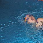
- severe load on the shoulder when turning inward, especially if the load is regular. This problem, for example, often occurs in crawl swimmers;
- Shoulder injuries of various nature can also be the cause.
About problems
What problems may arise when it comes to this part? human body? The first problem is subscapularis tendinopathy. These are some that arise as a result of insufficient blood supply to the muscles. The cause of tendinopathy can also be hereditary pathologies of connective tissue. It should be noted that, for example, with tendinopathy of the subscapularis muscle, a person’s pain intensifies when he brings a spoon to his mouth, combs his hair, or moves his arm behind his back. It should be noted that if a patient often has similar problems relating to the muscles of the shoulder girdle, then he may be diagnosed with “humeral periarthritis”. This problem can also arise in case of permanent tendon injury.

About breaks
Rupture of the subscapularis muscle does not occur very often. However, the cause most often is tendinopathy, which arises as a precursor to this problem. Symptoms of a subscapularis tendon rupture:
- sudden increase in pain;
- inability to move your arm freely.
It is also important to note that ruptures can be either partial or complete, where the tendon is completely torn from its attachment. The intensity of pain, of course, depends on this. With partial ruptures, movement of the hand may still be preserved (although it will be accompanied by pain), while with complete ruptures the hand is completely immobilized.
Diagnostics
Only a doctor can diagnose the problem. This will require examination of the patient and collection of anamnesis. The patient will also be sent for an x-ray. To clarify the extent of damage, you will have to undergo an ultrasound examination or MRI.

Treatment
How is the subscapularis muscle treated? Most often it is conservative. The main thing with a partial tendon rupture is to relieve the pain. For this purpose, painkillers and anti-inflammatory drugs may be prescribed. Also, the sore spot is fixed with a tight bandage. If a tendon ruptures, a splint may be used. When the pain goes away and the shoulder returns to normal, doctors prescribe special exercises to develop the joint.
Sometimes surgery is required. This is most often necessary in cases of complete tendon rupture, or if conservative treatment has not produced the desired results.
Injuries to the subscapularis tendon often go undetected, especially partial or full-thickness injuries of the superior portion of the subscapularis tendon. The incidence of these injuries is 27%. Many of them are combined with other injuries, especially rupture of the supraspinatus tendon
Repair of the subscapularis tendon plays a role important role in maintaining a balance of forces that ensures a stable axis of rotation during movements in the shoulder joint, since this tendon acts as a dynamic stabilizer, providing an anterior moment of the transverse pair of forces.
The mechanism of injury is forced external rotation, hyperextension.
Patient complaints
The main complaint of the patient with damage to the subscapularis tendon is pain. Many patients may deny an episode of injury, especially with degenerative or compound ruptures of the subscapularis tendon. Some patients cannot move their arm to its original position due to pain and limited movement.
Clinical examination
Clinical signs of subscapularis tendon injury include painful arching, decreased internal rotation strength, and increased passive external rotation.
To determine the integrity of the subscapularis tendon, special tests are performed:
- Lift-of test– assesses the patient’s ability to lift the arm when the dorsum of the hand is pressed against the lumbar region with maximum internal rotation (i.e., the function of the subscapularis muscle).
- Test of pressing your hand to your stomach (“belly-press” test)– the patient presses his hand to the anterior wall of the abdomen and moves the elbow forward to a horizontal position, using internal rotation. It is considered positive when the patient's elbow deviates posteriorly, and the pressure of the hand on the anterior abdominal wall is carried out due to shoulder extension.
- Napoleon test– is also performed, but taking into account the position of the wrist joint. In a patient, when the elbow deviates posteriorly, there is a corresponding flexion of the hand relative to the abdomen. This test may be negative if the damage is less than 50% (the hand is in a straightened state). If damage is more than 50%, but does not involve the entire tendon, an intermediate result can be obtained (i.e., flexion of the hand from 30° to 60°). Complete injury to the subscapularis tendon is indicated by a positive Napoleon test (i.e., wrist flexion to 90°).
- Bear hug test– the patient places the hand on the injured side on the opposite shoulder, keeping the elbow in a forward flexion position. The examiner attempts to lift the patient's hand from the shoulder with external rotation, which the subject counters with internal rotation. The test is considered positive if the examiner can lift the arm away from the patient's shoulder, indicating at least partial damage to the subscapularis tendon.
In addition, the presence of subcoracoid impingement, which may be an etiological factor and cause of pain in the shoulder joint, should be assessed.
A specialimpingement test, which is considered positive when pain and/or clicking occurs during passive anterior flexion, abduction, and internal rotation. Most patients note pain in the area of the coracoid process and subcoracoid zone.
X-ray:
- Axillary view - may indicate static anterior subluxation of the humeral head in patients with chronic subscapularis tendon failure.
- Anteroposterior projection - migration of the humeral head in the proximal direction is highly likely to indicate extensive damage to the rotator cuff, which may also involve the subscapularis tendon.
Ultrasonographyhas a confidence level of up to 85% when diagnosing full-thickness injuries of the subscapularis tendon, however, for partial ruptures and small injuries, the reliability of the method has not been confirmed.
When assessing rotator cuff pathology, the method of choice isMRI. Additional contrast allows better differentiation of pathological changes in the rotator cuff and can increase diagnostic confidence, especially for injuries of the upper parts of the subscapularis tendon. However, full-thickness injuries, superior tears, and partial subscapularis tendon injuries often go unrecognized on MRI (31% detection rate).
CT scan:
Axillary projection - the length of the apex of the coracoid process is determined (the normal length of the protruding part of the coracoid process is 8.2 mm). Excessive protrusion predisposes to subcoracoid impingement.
A more common measurement method is to determine the distance between the tip of the coracoid and the humerus/lesser tuberosity on CT and MRI images. Normally, the coracobrachial distance ranges from 8.7 to 11.0 mm. A decrease in this interval is associated with damage to the supraspinatus tendon; diagnosis of damage to the subscapularis tendon is difficult.
Treatment of subscapularis tendon injury
Conservative treatmentsubscapularis tendon injury generally corresponds to the treatment of posterosuperior rotator cuff tears and is used only for minor injuries in mature and elderly patients.


General principles of conservative treatment include:
- Cooling/warming treatments
- Taking painkillers, NSAIDs, injections (for example, subacromial or into the cavity of the shoulder joint)
- Physiotherapy
Surgical treatment:
The main method of treating this pathology isarthroscopic repair of the subscapularis tendon
Candidates for surgical intervention in the group of mature and elderly patients are people who, despite conservative measures carried out for 3-6 months, continue to have severe pain and dysfunction. When early diagnosis of damage to the subscapularis tendon of any degree of damage, it is advisable to immediately apply surgical treatment.
Main stages of the operationanchor fixation of the subscapularis tendonpresented in photographs:

1) Stage of diagnosing the degree and depth of damage

2) Treatment of the damaged area with an electrode and preparation of the defect area


4) Installation of an anchor from the area of tendon attachment to the lesser tubercle of the humerus

5) Suturing using any technique

6) Tightening the threads. Final fixation
Video on surgical technique for grade 2 subscapularis tendon injury.
Rehabilitation after injury to the subscapularis tendon
In the postoperative period, immobilization with a support bandage is recommended for 6 months while walking and sleeping. Strength exercises for the rotator cuff and deltoid muscles can be started by 12 weeks postoperatively.
The so-called frozen shoulder syndrome, accompanied by pain and limited mobility in the shoulder joint, can be the result of various injuries and disorders. Often this problem occurs due to overstrain of the subscapularis muscle. That is why in this article the site will tell you how to localize trigger points of the subscapularis muscle, what symptoms are characteristic of their appearance, as well as ways to eliminate pain caused by muscle strain.
Subscapularis muscle: location and main functions
The very name of the subscapularis muscle ( m. subscapularis) indicates its location - the muscle is located under the scapula, between the rib cage and the scapula bone. The muscle passes laterally and is attached to the anterior part of the humeral head of the shoulder joint.
When the subscapularis muscle contracts, the arm rotates inward and is brought toward the body. As one of the rotator cuff muscles, the subscapularis muscle supports the shoulder joint during movement, especially when the humeral head tends to slide upward during abduction.
Below we will look at:
- subscapularis trigger points and referred pain patterns;
- reasons for the appearance of trigger points of the subscapularis muscle;
- subscapularis trigger point symptoms;
- self-massage of the subscapularis muscle to eliminate pain.
As one of the rotator cuff muscles, the subscapularis muscle supports the shoulder joint during movement.
Subscapularis Trigger Points and Referred Pain Patterns
The subscapularis muscle can become the site of up to three trigger points, two of which usually appear near the outer edge of the muscle. Fortunately, a trigger point on the inner edge of the muscle is much less common because it is almost impossible to palpate and eliminate manually.

The main symptoms that accompany the presence of trigger points in the subscapularis muscle
The activity of trigger points in the subscapularis muscle can lead to the following symptoms and complaints:
- severe pain in the back of the shoulder, which can appear during movement or immobility of the shoulder and is localized quite deeply;
- inability to raise the arm more than 45 degrees - severe pain occurs when trying to raise the arm to the side;
- pain intensifies when trying to touch the armpit located under the other arm;
- frozen shoulder syndrome, or adhesive capsulitis: pain in the shoulder area, limited mobility in the shoulder joint.
Pain in the shoulder, under the shoulder blade, in the upper arm and around the wrist are possible symptoms of a strained subscapularis muscle.
A quick guide to self-massage of the subscapularis muscle
To massage the subscapularis muscle, it must first be localized. To do this, sit in a chair or lean forward, lower one arm down and relax it so that it dangles freely.
Place your other hand in the armpit and begin to rotate your shoulder inward - during this movement you will feel the subscapularis muscle. Remember that this area is very sensitive because it contains a large number of nerves. Feel the muscle contract, then relax it and begin the massage.
Make sure you only massage the muscle and not the nerves, as this can lead to armpit pain. The subscapularis muscle should be massaged thumb, slowly moving it over sensitive points of the muscle.
Another massage option is a pressure movement: apply pressure with your right hand to the muscle located on the left side, and then perform various movements with your left hand (lift, rotate it).
Similar articles
 From the experience of working as a teacher-psychologist in a summer health camp for schoolchildren Psychotherapeutic games for teenagers
 Tibetan sleep and dream yoga download fb2 Daytime sleep yoga practice: four steps in reality
 Recipes from the Atlas of Tibetan Medicine
 Russian team at international competitions in underwater robotics Competitions in underwater